Diseases of seabass, Lates calcarifer, larvae from hatcheries in Indonesia
| dc.contributor.author | Zafran | |
| dc.contributor.author | Koesharyani, Isti | |
| dc.contributor.author | Yuasa, Kei | |
| dc.contributor.author | Hatai, Kishio | |
| dc.contributor.editor | Lavilla-Pitogo, Celia R. | |
| dc.contributor.editor | Cruz-Lacierda, Erlinda R. | |
| dc.date.accessioned | 2021-11-09T15:53:29Z | |
| dc.date.available | 2021-11-09T15:53:29Z | |
| dc.date.issued | 2002 | |
| dc.identifier.citation | Zafran, Koesharyani, I., Yuasa, K., & Hatai, K. (2002). Diseases of seabass, Lates calcarifer, larvae from hatcheries in Indonesia. In C. R. Lavilla-Pitogo & E. R. Cruz-Lacierda (Eds.), Diseases in Asian aquaculture IV: Proceedings of the Fourth Symposium on Diseases in Asian Aquaculture, 22-26 November 1999, Cebu City, Philippines (pp. 279-284). Fish Health Section, Asian Fisheries Society. | en |
| dc.identifier.isbn | 9718020160 | |
| dc.identifier.uri | http://hdl.handle.net/10862/6219 | |
| dc.description.abstract | This paper describes three diseases of seabass (Lates calcarifer) larvae: viral nervous necrosis (VNN), glugeosis microsporidian infection, and a bacterial infection observed in private hatcheries in Indonesia from 1997-1998. VNN of seabass larvae was initially recorded in one hatchery in Situbondo, East Java in August 1997. Occurence in hatcheries in Bali and Banyuwangi, East Java was recorded in the same year. Cumulative mortalities in each case reached 100% within one week. The signs include abnormal swimming, or fish remaining on the tank bottom. Histopathologically, necrosis and vacuolation were observed in the brain and retina of affected larvae. Abundant spherical viral particles, 30 nm in diameter, were found in the cytoplasm of affected nerve cells. Glugeosis and bacterial infection were observed in seabass larvae in Banyuwangi, East Java in 1998. Larvae with glugeosis had numerous whitish cysts (xenoma), 0.5-1.5 mm in diameter, in the abdominal cavity. The cysts were composed of Glugea spores measuring 5-6.5 µm x 2.0-2.5 µm in size. The disease caused mortalities up to 10%. Outbreaks of bacterial disease resulted in 100% mortalities within one week. Diseased larvae showed sluggish swimming near the water surface or weak swimming near the tank bottom. Histological sections of liver tissues showed Grain-negative rods, but isolation of the bacteria was not successful. | en |
| dc.publisher | Fish Health Section, Asian Fisheries Society | en |
| dc.subject | Lates calcarifer | en |
| dc.subject | Indonesia | en |
| dc.subject | Glugeosis | en |
| dc.title | Diseases of seabass, Lates calcarifer, larvae from hatcheries in Indonesia | en |
| dc.type | Conference paper | en |
| dc.citation.spage | 279 | en |
| dc.citation.epage | 284 | en |
| dc.citation.conferenceTitle | Diseases in Asian aquaculture IV: Proceedings of the Fourth Symposium on Diseases in Asian Aquaculture, 22-26 November 1999, Cebu City, Philippines | en |
| dc.subject.asfa | fish diseases | en |
| dc.subject.asfa | sea bass culture | en |
| dc.subject.asfa | hatcheries | en |
| dc.subject.asfa | parasitology | en |
| dc.subject.asfa | bacteriology | en |
| dc.subject.asfa | histology | en |
| dc.subject.asfa | polymerase chain reaction | en |
| dc.subject.scientificName | Lates calcarifer | en |
Files in this item
| Files | Size | Format | View |
|---|---|---|---|
|
There are no files associated with this item. |
|||
This item appears in the following Collection(s)
-
Diseases in Asian aquaculture IV [43]
Proceedings of the Fourth Symposium on Diseases in Asian Aquaculture, 22-26 November 1999, Cebu City, Philippines
Related items
Showing items related by title, author, creator and subject.
-
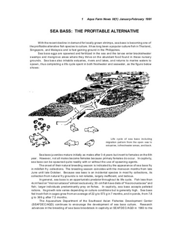
Sea bass: The profitable alternative
(Aquaculture Department, Southeast Asian Fisheries Development Center, 1991) -
Seabass grow-out and marketing: lessons from Australia, Malaysia, and Thailand
(Aquaculture Department, Southeast Asian Fisheries Development Center, 1997) -
A feed for seabass
(Aquaculture Department, Southeast Asian Fisheries Development Center, 1997)One of the major constraints in seabass (Lates calcarifer) culture is feed supply. Details are given of work conducted at AQD regarding the formulation of a 'standard' feed suitable for carnivorous species like the seabass and groupers. Diet formulae for seabass grow-out and for larval rearing are given.