Growth regulation by insulin-like growth factor-I in fish

Share
Abstract
Insulin-like growth factor-I (IGF-I) is a mitogenic polypeptide that plays an essential role in the regulation of development and somatic growth of vertebrates, mainly by mediating growth hormone actions. It has clearly been established that the structure of IGF-I and its biological function has been highly conserved among vertebrates. In this paper, we review the recent developments in the molecular, biochemical, and physiological properties of IGF-I in fish.
Suggested Citation
Moriyama, S., Ayson, F. G., & Kawauchi, H. (2000). Growth regulation by insulin-like growth factor-I in fish. Bioscience, Biotechnology and Biochemistry , 64(8), 1553-1562. https://doi.org/10.1271/bbb.64.1553
Type
Article; ReviewISSN
0916-8451Collections
- Journal Articles [1266]
Related items
Showing items related by title, author, creator and subject.
-
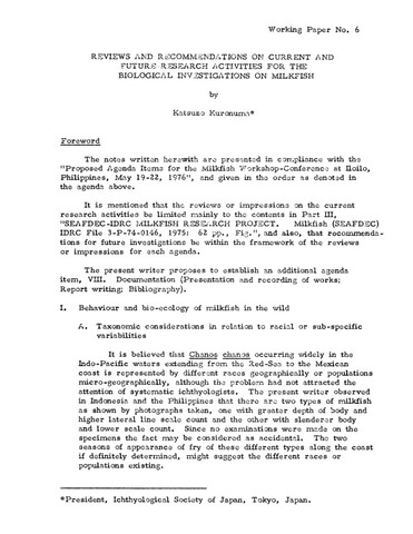
Reviews and recommendations on current and future research activities for the biological investigations on milkfish
(Aquaculture Department, Southeast Asian Fisheries Development Center, 1976)Presented in this paper are the recommendations on current and future research activities for the biological investigations on milkfish (Chanos chanos). Included were the bahavior and bio-ecology of milkfish in the wild. It also tackled the protocols in transporting broodstocks. -
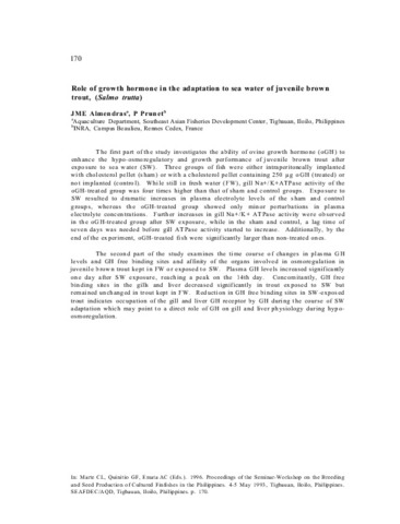
Role of growth hormone in the adaptation to sea water of juveline brown trout, (Salmo trutta)
(Aquaculture Department, Southeast Asian Fisheries Development Center, 1996)The first part of the study investigates the ability of ovine growth hormone (oGH) to enhance the hypo-osmoregulatory and growth performance of juvenile brown trout after exposure to sea water (SW). Three groups of fish were either intraperitoneally implanted with cholesterol pellet (sham) or with a cholesterol pellet containing 250 µg oGH (treated) or not implanted (control). While still in fresh water (FW), gill Na+/K+ATPase activity of the oGH-treated group was four times higher than that of sham and control groups. Exposure to SW resulted to dramatic increases in plasma electrolyte levels of the sham and control groups, whereas the oGH-treated group showed only minor perturbations in plasma electrolyte concentrations. Further increases in gill Na+/K+ ATPase activity were observed in the oGH-treated group after SW exposure, while in the sham and control, a lag time of seven days was needed before gill ATPase activity started to increase. Additionally, by the end of the experiment, oGH-treated fish were significantly larger than non-treated ones. The second part of the study examines the time course of changes in plasma GH levels and GH free binding sites and affinity of the organs involved in osmoregulation in juvenile brown trout kept in FW or exposed to SW. Plasma GH levels increased significantly one day after SW exposure, reaching a peak on the 14th day. Concomitantly, GH free binding sites in the gills and liver decreased significantly in trout exposed to SW but remained unchanged in trout kept in FW. Reduction in GH free binding sites in SW-exposed trout indicates occupation of the gill and liver GH receptor by GH during the course of SW adaptation which may point to a direct role of GH on gill and liver physiology during hypo-osmoregulation. The second part of the study examines the time course of changes in plasma GH levels and GH free binding sites and affinity of the organs involved in osmoregulation in juvenile brown trout kept in FW or exposed to SW. Plasma GH levels increased significantly one day after SW exposure, reaching a peak on the 14th day. Concomitantly, GH free binding sites in the gills and liver decreased significantly in trout exposed to SW but remained unchanged in trout kept in FW. Reduction in GH free binding sites in SW-exposed trout indicates occupation of the gill and liver GH receptor by GH during the course of SW adaptation which may point to a direct role of GH on gill and liver physiology during hypo-osmoregulation. -
Growth response of cultured larvae of silver therapon Leiopotherapon plumbeus (Kner, 1864) in outdoor tanks in relation to fertilizer type and fish density
Aya, Frolan ; Garcia, Luis Maria (Wiley, 2016)
This study evaluated the effects of fertilizer type and fish density on early growth and survival of silver therapon Leiopotherapon plumbeus (Kner, 1864) larvae reared in outdoor tanks. In the first experiment, larvae (1.92 ± 0.09 mm total length) were stocked into nine, 4 m3 tanks at an initial density of 0.5 larvae L-1 and reared for 42 days at an ambient temperature of 28.8–30.7°C. Three treatments with three replicates each were compared: organic (chicken manure, OF) or inorganic fertilizers (ammonium phosphate, IF) applied once every 2 weeks, and the unfertilized (NF) tanks serving as the control group. Water quality, zooplankton densities, survival or growth of L. plumbeus larvae did not vary significantly in either fertilized or unfertilized tanks. Fertilization resulted in elevated nutrient concentrations, which did affect survival (2.10%–6.07%) of the fish larvae. In the second experiment, larvae were stocked at densities of 0.4 or 0.6 larvae L-1 in tanks fertilized at 4–5 days interval with OF and IF for 30 days. Growth performance of L. plumbeus larvae was affected by fish density, with significantly larger (20.04 ± 2.65 mm in total length) and higher specific growth rate (SGR; 6.97 ± 0.48% day-1) at 0.4 larvae L-1 than at 0.6 L-1. Fry production did not vary significantly between fish density treatment groups given the same fertilizer types, but survival rates were improved at 0.4 L-1. Together, production of L. plumbeus larvae in outdoor tanks can be optimized at a lower stocking density, regardless of the type of fertilizer used.
; Garcia, Luis Maria (Wiley, 2016)
This study evaluated the effects of fertilizer type and fish density on early growth and survival of silver therapon Leiopotherapon plumbeus (Kner, 1864) larvae reared in outdoor tanks. In the first experiment, larvae (1.92 ± 0.09 mm total length) were stocked into nine, 4 m3 tanks at an initial density of 0.5 larvae L-1 and reared for 42 days at an ambient temperature of 28.8–30.7°C. Three treatments with three replicates each were compared: organic (chicken manure, OF) or inorganic fertilizers (ammonium phosphate, IF) applied once every 2 weeks, and the unfertilized (NF) tanks serving as the control group. Water quality, zooplankton densities, survival or growth of L. plumbeus larvae did not vary significantly in either fertilized or unfertilized tanks. Fertilization resulted in elevated nutrient concentrations, which did affect survival (2.10%–6.07%) of the fish larvae. In the second experiment, larvae were stocked at densities of 0.4 or 0.6 larvae L-1 in tanks fertilized at 4–5 days interval with OF and IF for 30 days. Growth performance of L. plumbeus larvae was affected by fish density, with significantly larger (20.04 ± 2.65 mm in total length) and higher specific growth rate (SGR; 6.97 ± 0.48% day-1) at 0.4 larvae L-1 than at 0.6 L-1. Fry production did not vary significantly between fish density treatment groups given the same fertilizer types, but survival rates were improved at 0.4 L-1. Together, production of L. plumbeus larvae in outdoor tanks can be optimized at a lower stocking density, regardless of the type of fertilizer used.